Yeah, I know. Sorry about that headline. Being a Physiologist, one might think me biased, but Physiology is, of course, the most fascinating of all sciences. Some people might disagree with that, but they are wrong. Within the broad subject of Physiology, there are certain areas that are even more interesting than others. Opinions may vary on which aspects of Physiology are the most fascinating, but pretty much everyone would put the nervous system at or near the top of the list, and within the subject of the nervous system, most people find the organs of the “special senses” to be pretty amazing. Special senses refers to sensory inputs to the nervous system that rely on highly specialized sense organs—vision is one of the special senses, because it is mediated by the eyes. Hearing and balance (which rely on the ears and the (also very cool) organs of the inner ear, smell (the olfactory apparatus) and taste (taste buds) are the other special senses.
We’ve already talked a bit about the nature of light. Visible light is a very small part of the spectrum of electromagnetic energy that is detectable by our eyes. The energy would be there, whether we saw it or not, just like ultraviolet radiation from the sun, radio waves or x-rays. These are all also forms of electromagnetic energy, like visible light, but we can’t detect them directly with any of our senses. If our eyes were “tuned” a little differently, we could see infrared radiation or UV, like many other creatures can, but we are limited to seeing those wavelengths of electromagnetic energy that we detect as light that ranges from red light on one end of the visible spectrum to violet on the other. That’s why “infrared” is called “infrared”. The word simply means “below the red”, because it is the wavelengths of electromagnetic energy just a bit longer than the wavelengths we see as red light. Similarly, “ultraviolet” means “beyond violet”, and describes the wavelengths just a little shorter than those that we see as violet light. If you Google “images for electromagnetic spectrum”, this will be a little more clear, since pictures (pictures being reflected light you see with your eyes, by the way) are worth a thousand words.
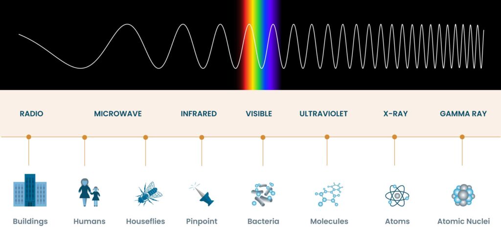
Source: NASA
So, we can see the spectrum of visible light between red and violet. The process of vision is, as you might imagine, complex. It only starts with the eyes. It’s actually a part of your brain, at the back of the brain, called the visual cortex that takes the light your eyes collect and turns it into what you perceive as vision. By the way, one of the reasons you “see stars” when you take a blow to the back of the head is because the impact causes the visual cortex to react, and you “see” flashes of light, as a result.
Electromagnetic energy is not something your brain can detect, directly, just as sound waves aren’t detectable by your brain, either. Your eyes are receptors for electromagnetic energy, and they are among the most amazing of all anatomical structures. An eyeball is, for all intents and purposes, an optical instrument, much like a microscope or telescope. All of these function is to gather light and focus it so that you can see it.

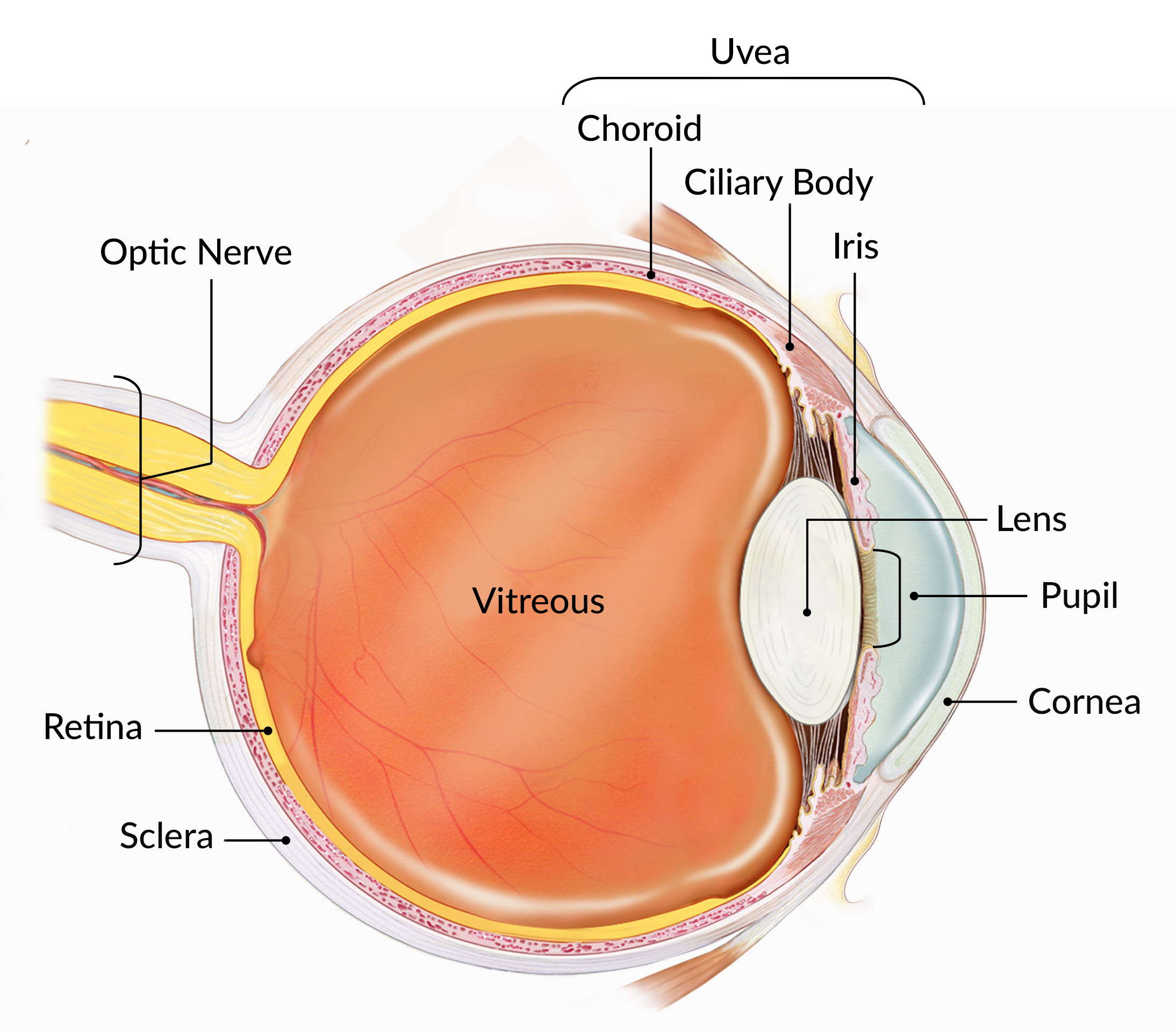
Source: National Eye Institute, National Institutes of Health
Light energy first enters the eye and is focused on to your retina by some of the most unique tissues in your body—the cornea (the clear outside covering of your eye) and the lens which is also a clear structure that sits just behind your iris, which is the pretty, colored part. The pupil is the hole in the iris through which light passes. The iris is actually a muscle that can contract or relax. As it contracts, the pupil gets smaller, which allows less light to enter the eye, as when it’s bright and sunny. Too much light can damage the photoreceptors in the retina. When it’s darker, the iris relaxes, the pupil gets larger and more light can enter the eye, allowing you to see better. The contraction of the iris is controlled by a reflex, in response to light. This reflex is what your doctor is looking for when they do that thing where they shine that little pen light in your eyes. If you watch medical shows on TV, you’ve probably heard things like, “the pupils are fixed and dilated” when someone is dead or has suffered a brain injury. This happens because they irises no longer respond to the light and relax, dilating the pupils. Sometimes, one eye will respond and the other won’t, indicating that one side of the brain has been damaged. Some drugs also interfere with the pupil response, so a person experiencing an overdose may sometimes have “pinhole” pupils because their irises are fully contracted, regardless of light.
I say the cornea and lens are particularly amazing tissues for a couple of reasons. The big reason is that they are both clear (at least in young eyes). The cornea and lens are both living tissues composed mostly of cells and proteins. How are they clear, then? That’s a really good question with a really complicated answer that I only understand a bit. First off, you need to understand that, despite all the pictures you may have seen in books, most cells are, actually, mostly clear—sacs of fluid with some stuff here and there inside. In fact, if you just put a cell under a microscope, you generally can’t see much until you apply a stain to the cell that allows you to see the structures. You also need to get a handle on what “clear” means. Whenever we see something, it is because that thing has reflected light back to our eyes. A leaf on a tree, for instance, reflects green light, which we see. The light from the sun is what is called “white light”, meaning it is a mixture of all the different wavelengths (colors) of light. The leaf reflects the green light and absorbs the other colors. Remember the article about fall colors in leaves? It is the energy in the non-green colors that the leaf absorbs and that the plant uses to harvest energy. The green isn’t absorbed—it bounces off (reflects) and that is the light we see. When something is clear, it allows light to pass through—it neither reflects or absorbs the light. When you see cells, it’s because they contain pigments or other things inside the cells that reflect light. Otherwise, they are clear. Now, outside of the cornea and lens, the rest of our tissues are obviously not clear. The reason for that is mostly because of blood and blood vessels and the fact that some cells, like muscle cells also contain a lot of proteins. The oxygen and nutrients that most all of our cells need is delivered in the blood, so there is a lot of blood and a lot of blood vessels to carry it. The cells of the cornea and lens, however, get their nutrition and oxygen from the clear fluids that fill the eye and the tears that lubricate the outside layer of the cornea. There are actually special biochemical factors produced in the cornea and lens that prevent blood vessels from growing in the tissues and messing up the clarity. The blood vessels you see when your eyes are “bloodshot” aren’t in the cornea—they are in the sclera, which is the white tissue under the cornea.
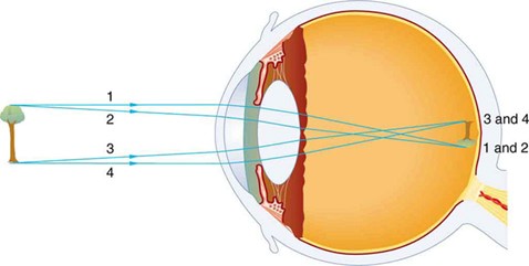
Source: University of Tulsa
Anyway, light enters the eye, passing through the cornea, through the pupil, and the lens. The cornea and lens act like the lenses on a camera to focus the light onto the back of the eye, just like focusing a camera to cause the image to fall very clearly on the film (or the digital photoreceptor in a digital camera). The lens is flexible and is attached to a muscle, all around the edges. When the muscle contracts, it pulls on the edges of the lens, causing the lens to stretch, which makes it thinner. When the muscle relaxes, the tension in the lens decreases and the lens gets thicker. The shape of the lens determines where the light will be focused. As we age, the lens tends to get stiffer. It still stretches (which allows us to focus on things farther away), but it doesn’t relax and get thicker as well as it did when we were young, which makes it harder to focus on things close up. This is why older people usually need reading glasses. The structure at the back of the eye is called the retina, and it is where the actual photoreceptors of you eye are located. This is like the film in a camera. The retina is very densely packed with nerve cells and blood vessels. Some of the nerve cells are specialized to react to light. These are called photoreceptors. We have two kinds of photoreceptors in our retinas. One type, referred to as “rods”, is sensitive to varying intensities of light, but not to color. The other type of photoreceptors are called “cones”. We have three different types of cones. One type is sensitive to red light, one type is sensitive to blue light and one type is sensitive to green light. You can combine those three colors, red, blue, and green, in differing ratios and get any possible color. That is how color TV’s work. Each pixel has red, blue and green and depending on what color that particular pixel is supposed to be to make the picture, varying amounts of red, blue and green will be activated. Your eyes work the same way. If you see something purple, the purple light will activate some blue cones and some red cones, which your brain will later interpret as purple. Your rods are much more sensitive in low light than are your cones, which is why your vision is mostly black-and-white when it’s dark. In very low light, only the rods are activated, because the cones need more light to work.
The rods and cones are packed together in the retina, so when an image, like that tree leaf, is focused on the retina, the photoreceptors that the image hits are activated. Because the image is green, the green cones will be particularly activated, although the others will be to, so that you can see shades of colors, brightness and so on. When activated, the rods and cones convert the light energy into electrical impulses that leave your eye through your optic nerve, pass into specific pathways in your brain and are transmitted to the visual cortex, located at the back part of your brain. The visual cortex knows what each electrical signal it receives means, in terms of color, brightness and location, so your visual cortex forms a “map” of the image that was focused on your retina and then your brain tells you what that image means. There is a lot of other stuff that goes on with how visual input is processed and interpreted by your brain, like how we see movement, but that is probably more than we should get into now. How you see movement is so cool, it’s hard to believe how much brain power is used to interpret it.
There are other aspects of sight to touch on. We have two eyes, with a big honking nose in between them. Why two eyes and why do we only see one image, with no nose in the way in the middle? We have two eyes so that we can judge distance. When you look at that leaf, an image is seen in both eyes, but from slightly different angles. You brain is so good and interpreting the images your eyes receive that it will take those two, slightly different images and be able to tell how far away the leaf is (relatively speaking). If the leaf is close to your eyes, the difference in the angle between your right eye and your left eye will be quite a bit, and your brain will interpret that as “close”. If the leaf is far away, the angle is less and your brain sees that as “far”. Some people are better at judging that angle and those people have what we refer to as better “depth perception”. Generally, people whose eyes are farther apart will have better depth perception than people whose eyes are close together, because when the eyes are farther apart, the angle is relatively larger so your brain gets better information. That’s why people who lose an eye have problems with depth perception. They can still judge distance, somewhat, but they have to rely on visual clues like comparing sizes of things and perspective (things further away look smaller, etc.)
The reason why you only see one image and why you don’t see your nose in the middle is that your brain combines the information from both eyes into a single image and it also “edits out” your nose and fills in the hole in the image by taking information from both eyes and interpreting what your nose was blocking out. It is a staggeringly complex and awesome physiological process. Another really amazing aspect of all of that is that, in order to function properly, both of your eyes need to be pointed in the same direction. EXACTLY the same direction. The muscles that control the movement of your eyes are tiny, but there is a very large part of your brain dedicated to controlling them and making them track together. Try this experiment—go look at yourself in a mirror. Stare into your eyes. Now move your head. Your eyes will stay perfectly locked in place. No matter how fast you move and shake your head, your eyes will remain perfectly still, staring straight back at you. If you think about how complicated that is and how fast your eyes can move (because as you move your head in the mirror, your eyes are actually moving to stay locked in), it is mind-boggling. I keep telling you, science is some really cool stuff. Just in this little article, we went, not just into Physiology, but into Biology (all the cells, blood and stuff), Chemistry (the molecules and chemical signals) and Physics (the optics of vision, electromagnetism, RGB additive colors). It’s always all tied together and life just gets more interesting the more you know and the more you wonder about things.